
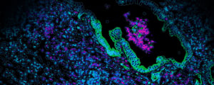
Spatial Proteomics & Multiomics
Beyond gene expression. Protein expression in spatial context.
Explore proteins, cellular interactions and tissue biology in spatial context.
Spatial proteomics enables researchers to investigate protein expression within intact tissues, preserving the cellular organisation and microenvironment that are lost through conventional dissociation-based approaches.
At the South Australian Genomics Centre (SAGC), we provide access to advanced spatial proteomics and multiomics technologies, including CosMx Spatial Molecular Imager, 10x Genomics Xenium and STOmics Stereo-CITE, supporting high-resolution protein profiling and integrated RNA–protein analysis.From tumour immunology to neuroscience and developmental biology, these technologies enable researchers to investigate cellular phenotypes, functional states and molecular interactions in unprecedented spatial detail.
Why Spatial Proteomics?
While spatial transcriptomics reveals where genes are expressed, spatial proteomics provides complementary information about the proteins that define cellular identity, function and behaviour.
By measuring multiple protein markers directly within tissue sections, researchers can investigate complex cellular populations and understand how their spatial organisation contributes to biological processes.
Key advantages of spatial proteomics include:
- Identify cellular phenotypes: Characterise cell populations using multiple protein markers while preserving tissue architecture.
- Explore cellular interactions: Investigate how immune, stromal and other cell populations are organised within tissue microenvironments.
- Understand functional states: Measure proteins associated with signalling, activation, differentiation and disease processes.
- Investigate tissue heterogeneity: Reveal spatial differences in protein expression across tissue regions and cellular neighbourhoods.
- Integrate RNA and protein: Combine transcriptional and protein measurements for a more comprehensive understanding of cellular identity and function.
With high-plex spatial technologies, researchers can move beyond traditional immunohistochemistry and limited-marker immunofluorescence to investigate dozens or hundreds of molecular features within the same tissue section, depending on the assay.
EXPLORE OUR SPATIAL PLATFORMS
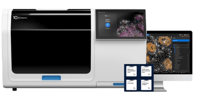
Xenium In Situ
Visualise selected RNA targets and protein markers within intact tissue to investigate cell types, cellular neighbourhoods and tissue organisation.
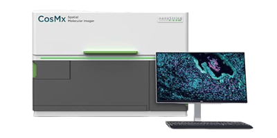
CosMx SMI
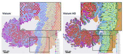
Visium HD
High-resolution transcriptome-wide discovery
Stereo-seqTM
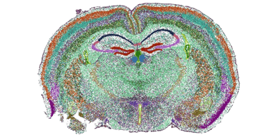
Whole-transcriptome spatial profiling at scale
Profile gene expression across diverse species and tissue types, including fresh-frozen and FFPE samples, with options for automated, large-scale studies.
EXPLORE OUR PROTEOMICS AND MULTIOMICS PLATFORMS
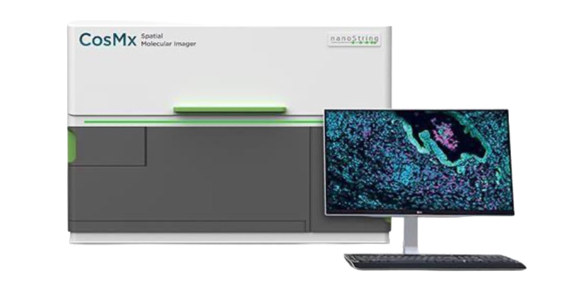
CosMx Spatial Molecular Imager (SMI)
HIGH-PLEX SPATIAL PROTEOMICS AT SINGLE-CELL RESOLUTION
The CosMx Spatial Molecular Imager enables highly multiplexed, imaging-based protein analysis within intact tissue sections, providing detailed information about cellular phenotypes, protein expression and tissue organisation.
Using oligonucleotide-tagged antibodies, CosMx supports the detection of dozens of proteins within the same FFPE tissue section, with spatial information at single-cell and subcellular resolution.
There are two ‘off-the-shelf’ specialised protein panels:
MOUSE NEUROSCIENCE PROTEIN PANEL
Designed for neuroscience research, this panel enables high-plex analysis of neural and glial cell populations, including markers associated with neuroinflammation, neurodegeneration and cellular function.
Applications include:
- Alzheimer's disease and neurodegeneration
- Microglial and astrocyte biology
- Neuroinflammation and immune responses
- Neural cell phenotyping
- Spatial organisation of brain tissue
HUMAN IMMUNO-ONCOLOGY PROTEIN PANEL
Designed to investigate the tumour microenvironment, this panel enables detailed characterisation of immune and tumour-associated cell populations, including markers associated with immune activation, checkpoint signalling and cellular phenotypes.
Applications include:
- Tumour immune microenvironment profiling
- Immune cell phenotyping and spatial organisation
- Immunotherapy and treatment-response research
- Biomarker discovery and validation
- Investigation of tumour–immune interactions
Integrated RNA and Protein Analysis
CosMx also supports spatial RNA profiling, enabling researchers to combine transcriptional and protein information within the same tissue section using compatible workflows. This integrated approach provides complementary insights into cellular identity, gene expression and protein-level functional states.
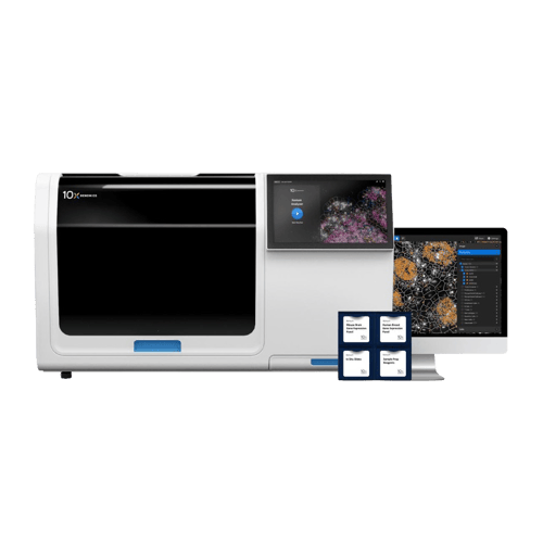
Xenium In Situ — Spatial RNA & Protein
TARGETED MULTIOMIC PROFILING AT SUBCELLULAR RESOLUTION
The 10x Genomics Xenium platform combines high-resolution spatial gene expression profiling with complementary protein detection, enabling researchers to investigate RNA and protein expression within the same tissue section.
Using targeted RNA panels alongside validated protein markers, Xenium supports detailed characterisation of cellular populations, immune phenotypes and tissue microenvironments.
This approach is particularly valuable when researchers have established molecular targets of interest and want to investigate how RNA and protein expression relate to cellular identity and tissue organisation.
Key applications include:
- Immune cell characterisation and phenotyping
- Tumour microenvironment analysis
- Investigation of immune checkpoint markers
- Spatial biomarker validation
- Integrated RNA and protein analysis in FFPE tissues
Xenium provides a targeted multiomic approach that complements the broader protein profiling capabilities available through CosMx.
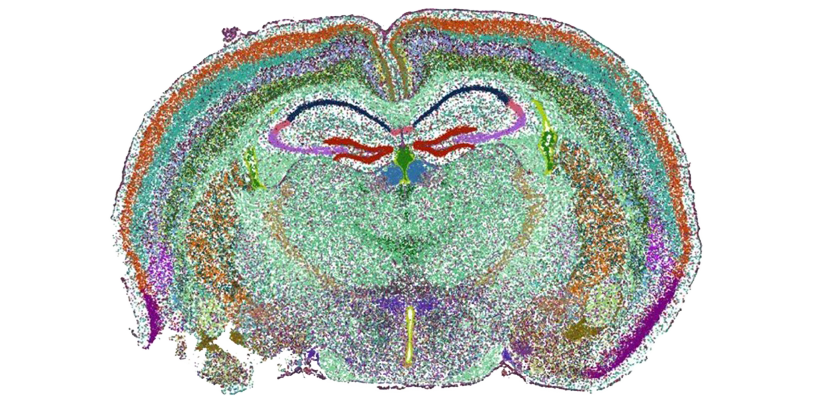
Stereo-CITE — Whole-Transcriptome RNA & Protein
UNBIASED SPATIAL TRANSCRIPTOMICS MEETS HIGH-PLEX PROTEIN PROFILING
STOmics Stereo-CITE combines whole-transcriptome spatial RNA profiling with high-plex protein detection, enabling researchers to investigate gene expression and selected protein markers within the same tissue section.
Unlike targeted RNA imaging approaches, Stereo-CITE provides transcriptome-wide gene expression information alongside antibody-based protein measurements, supporting integrated analysis of cellular identity, molecular pathways and tissue organisation.
With panels supporting up to 150 human or 120 mouse cell-surface protein markers, Stereo-CITE offers a powerful approach for investigating complex tissue environments.
Key advantages include:
- Whole-transcriptome spatial RNA discovery
- High-plex protein detection
- Integrated RNA and protein profiling from the same tissue section
- High-resolution mapping of cellular populations
- Analysis of cellular heterogeneity and spatial organisation
Stereo-CITE is particularly well suited to studies where researchers want the breadth of whole-transcriptome discovery combined with the additional biological information provided by protein markers.
Applications of Spatial Proteomics & Multiomics
Cancer Immunology and the Tumour Microenvironment
Investigate how immune cells, tumour cells and stromal populations interact within complex tissue environments.
Spatial protein profiling can help identify immune cell populations, investigate checkpoint marker expression and reveal how cellular organisation relates to tumour biology and treatment response.
Neuroscience and Neurodegenerative Disease
Characterise neural and glial cell populations within their anatomical context.
Spatial proteomics provides opportunities to investigate neuroinflammation, microglial activation, astrocyte responses and changes in protein expression associated with neurodegenerative disease.
Immunology and Inflammatory Disease
Explore immune cell diversity, activation states and spatial organisation across tissues.
Combining RNA and protein measurements can help distinguish immune cell populations and investigate molecular interactions that contribute to inflammation, tissue damage and immune regulation.
Biomarker Discovery and Validation
Investigate candidate biomarkers within their native tissue environment.
Spatial multiomics enables researchers to examine relationships between RNA expression, protein abundance and cellular localisation, supporting the investigation of disease-associated molecular signatures.
Developmental and Tissue Biology
Understand how cellular phenotypes and molecular programmes contribute to tissue development and organisation. Integrated RNA and protein profiling can reveal how transcriptional states relate to cellular differentiation, tissue architecture and developmental processes.
CHOOSING THE RIGHT SPATIAL PROTEOMICS APPROACH
Different technologies offer complementary strengths depending on the biological question, molecular targets and tissue requirements.
CosMX SMI
Primary strength
High-plex, protein-focused spatial
imaging
Best suited for
Immune phenotyping, tumour microenvironments, neuroscience
Xenium
Primary strength
Targeted RNA with complementary protein markers
Best suited for
Focused RNA–protein studies and biomarker characterisation
Stereo-CITE
Primary strength
Whole-transcriptome RNA with high-plex protein detection
Best suited for
Discovery-driven spatial multiomics and cellular heterogeneity
From Tissue to Biological Insight
SAGC supports researchers throughout the spatial proteomics and multiomics workflow, from experimental planning and tissue preparation through to data generation and downstream analysis.
Our services include:
- Experimental design: Platform selection, panel selection, sample numbers and study design.
- Tissue preparation and histology: Guidance on tissue compatibility, sectioning and quality control.
- Spatial molecular profiling: Access to CosMx, Xenium and Stereo-CITE workflows.
- Integrated data analysis: Cell type identification, spatial expression analysis, cellular neighbourhoods and RNA–protein integration, subject to platform and software compatibility.
- Bioinformatics support: Access to expertise and advanced spatial analysis solutions, including BioTuring SpatialX.
By bringing together complementary spatial technologies and analysis expertise, SAGC helps researchers move from complex molecular datasets to meaningful biological insights.
PLANNING A SPATIAL PROTEOMICS OR MULTIOMICS PROJECT?
Whether you're interested in high-plex protein profiling, investigating cellular interactions or integrating spatial RNA and protein measurements, our team can help you explore the available technologies and design an approach suited to your research.